 Image by Emily Finn
A new study published in the latest Nature Neuroscience journal used special "functional" MRI scans (f-MRI) to help determine why anorexia nervosa (AN) patients are so frightfully difficult to "cure" and and to embrace an effective treatment plan. Of all the mental disorders, AN is among the most -- if not the most -- prone to relapse and lethal outcomes, with over half of patients discharged in remission relapsing within one year.
The new study, done by researchers from New York State Psychiatric Institute at Columbia University Medical Center, and the NYU Dept. of Psychology and the New York State Psychiatric Institute, led by Drs. Joanna Steinglass and B. Timothy Walsh, studied 21 women with AN and compared their results using f-MRI with an equal number of healthy women while they made decisions about what foods to eat.
The anorexic women were more likely than the healthy women to choose low-fat, low-calorie foods, and they were less apt to perceive the high-fat, high-calorie foods as stimulating their food cravings.
Both AN patients and the controls showed activation in an area known as the ventral striatum, part of the brain s reward center. But the AN women showed more activity in the dorsal striatum, an area involved with habitual behavior. This suggested to the authors that the AN patients were acting automatically based on past learning, rather than weighing the pluses and minuses of the particular foods in front of them.
The main thrust of the authors' conclusions includes these considerations:
"This study examined the neural correlates of maladaptive food choices in individuals with anorexia nervosa. Prior neuroimaging studies of anorexia nervosa have examined the neural responses to passive viewing of food without examining the neurobiological mechanisms underlying active food choices. Our results suggest that a direct examination of food choice may be particularly useful for probing the neural basis of the persistent maladaptive behavior that is a salient characteristic of this disorder.
"Given that restrictive food intake is a persistent and clinically challenging problem in anorexia nervosa, these findings provide new insight into the neural mechanisms underlying this enigmatic illness," the authors continued. "Furthermore, as human and animal data have documented that the dorsal striatum has a critical role in the establishment and expression of action control and learned automatic behaviors, our results are consistent with the possibility that the persistent, maladaptive food choice in anorexia nervosa is subserved by fronto-striatal networks that are crucial for the development of habitual behavior.
In layman's terms, this means that f-MRI studies such as this one, show that AN patients' brain functions differ significantly from healthy people when contemplating food choices, and the specific locations of the brain activity detected among AN patients in the dorsal striatum region, as opposed to the more typical excitation of the "pleasure center" in the ventral striatum.
To take this a step further, this supports the rationale of habitual, ingrained behavior as a target for reducing the "maladaptive" (harmful, destructive) eating behavior so characteristic of AN, and explains why other approaches such as antidepressants and cognitive training don't work with AN. Instead, early intervention is necessary along with behavioral cues.
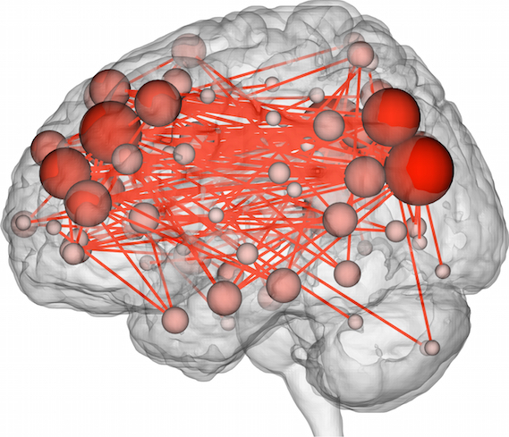
A related study in the same journal shows how idiosyncratic i.e., unique, "fingerprint"-like are our brain activity patterns on f-MRI. This study evaluated 126 patients via f-MRI and was able to pick out individuals based on their unique brain activity patterns. This one, entitled Functional connectome fingerprinting: identifying individuals using patterns of brain connectivity, was done by scientists at the Yale University Depts. of Neuroscience, Psychology and Radiology.