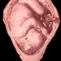
There is little as fascinating as maternal-fetal physiology, given that pregnancy itself is the most well-designed suspension system in the world. Add to this complexity the dynamic nature of birth. The transition required to be sustained by the umbilical cord inside the womb, then squeeze and pass through the vaginal canal into the outside world where the cord is cut and a baby takes his or her first breath. It is a continuous cascade of events that for mother and infant is no walk in the park, even under the most ideal conditions. Now, a study in PLoS ONE hopes to shed light on just how stressful an average, uneventful delivery is on the fetal head and brain - and it has the 3D magnetic resonance imaging (MRI) scans to show for it.
You see, the skull at that young age resembles plate tectonics. It is made of multiple soft pieces, that have some built-in compliance features like suture lines, spaces and general elasticity which morph and bend when compressed. This malleability facilitates undergoing the pressure and stress of delivery and afterwards affords the baby’s brain room to grow as he or she matures and develops. It is not until later that things fully fuse and the skull becomes more fixed. All of this enables a baby to be born and that is why the head often appears misshapen due to the changes it undergoes from the extent and duration of pressure it endures in the birth canal. This is called molding.
Sometimes the changes involve more extensive overlapping of the bony plates. This is what was depicted in the research just published where “3D MRI assessment using 3D finite element mesh reconstruction to compare the head between pre-labor and the second stage of labor” was performed. The sample size was very small and there were many limitations to the work given all the moving parts of delivery and ensuring the safety of mom and baby - but, the pictures are captivating, see here. That said, the researchers do raise the question of whether what we consider normal labor can be considered that when some fetal heads and brains undergo substantial deformation.
With the observations from these real-time simulations, they suggest “childbirth should perhaps be considered normal when a parturient gives birth naturally with only a few expulsive efforts, and her child is fine and has no consequences due to the shape changes of its brain during the process.” At present, a wide range of findings would fall under the “normal birth trauma” umbrella. For instance, it is not uncommon in an otherwise normal vaginal birth to have some bruising, even retinal or brain hemorrhages (or bleeds) and a fractured clavicle. This is often due to maternal and fetal factors (eg large fetal head, narrow maternal pelvis). This study only explored low risk scenarios, and only a few at that.
The utility of this imaging does inform beyond what can be observed with existing digital exams or external device monitoring. But, MRI is currently a cumbersome, impractical tool and how it can adapt for pathological births is unknown. Maybe that will change. As it stands, what was required to obtain this data warranted a significant care burden to ensure maternal and fetal safety in births that had low risk pregnancies, and still a couple needed C-sections. Understanding ways to “detect and prevent biomechanical risks linked to childbirth” is always a worthy endeavor and as a result of the ethical considerations necessary to evaluate real-time birth comprehensively the process will be complicated.
Source:
Ami O, Maran JC, Gabor P, Whitacre EB, Musset D, Dubray C, et al. (2019) Three-dimensional magnetic resonance imaging of fetal head molding and brain shape changes during the second stage of labor. PLoS ONE 14(5): e0215721. https://doi.org/10.1371/journal.pone.0215721